Lumbar Spinal Stenosis
Lumbar Spinal Stenosis
What is Lumbar Spinal Stenosis?
Symptoms
Treatment Methods
For those who do not experience improvement with conservative treatment, we recommend surgery.
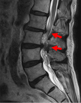
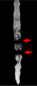
図1
Surgical Methods
Surgical methods are broadly divided into decompression surgery and fusion surgery. At our hospital, we strive to perform minimally invasive endoscopic surgery for decompression procedures whenever possible. Fusion surgery is indicated when there is instability or deformity, such as scoliosis, in the area of nerve compression. Fusion surgery is further divided into posterior surgery and anterior-posterior surgery, as outlined below.
- Minimally Invasive Posterior Lumbar Interbody Fusion (Mini-open TLIF + PPS)
This fusion surgery involves decompressing the spinal canal through a small midline incision, inserting an artificial spacer between the vertebrae, and then inserting screws through another small lateral incision.
- Minimally Invasive Anterior-Posterior Fusion (XLIF/OLIF + PPS)
This fusion surgery involves making a 5 cm incision in the side of the abdomen to insert an artificial spacer between the vertebrae, followed by screw insertion through a small incision in the back. One of the advantages of this method is that it indirectly decompresses the nerves without directly exposing them.
There are also surgeries that combine both procedures ① and ②. (See Figure 3)
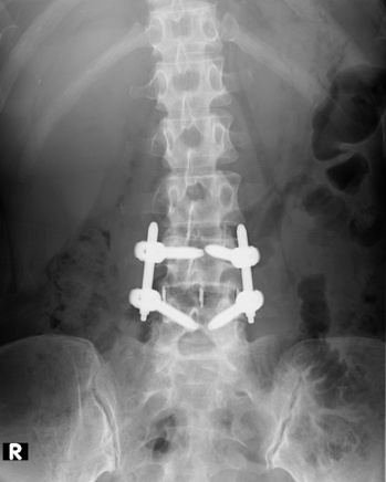

図2
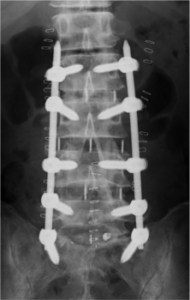
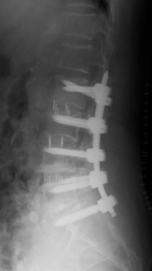
図3
Surgery Video
About Endoscopic Surgery
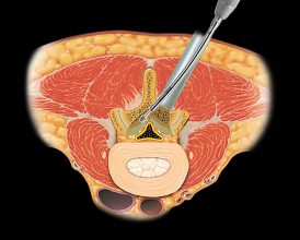
FESS (Full Endoscopic Spine Surgery)
Full endoscopic spine surgery is the least invasive endoscopic procedure, performed using an 8mm endoscope. By irrigating the surgical site with water from the camera, the procedure provides a clear view and allows for precise hemostasis.
-2-300x200.jpg)
MED, MEL (Micro Endoscopic Discectomy, Micro Endoscopic Laminectomy)
These endoscopic procedures, including micro endoscopic discectomy and laminectomy, are performed through an approximately 16mm incision. Our hospital has over 20 years of experience with these surgeries and has built a wealth of expertise.
BESS, UBE (Biportal Endoscopic Spine Surgery, Unilateral Biportal Endoscopic Surgery)
This is the latest endoscopic surgical technique, performed by making two incisions of approximately 5 to 8mm. A camera is inserted through one incision, with water irrigation similar to FESS, while surgical instruments are inserted through the other incision. In recent years, this method has become widely practiced, especially in Asia and the United States.
In some patients, the intervertebral disc may collapse, and bone deformation can lead to nerve compression. In such cases, spinal fusion surgery with screw insertion may be necessary.